Your cart is currently empty!
Furcation Repair Technique
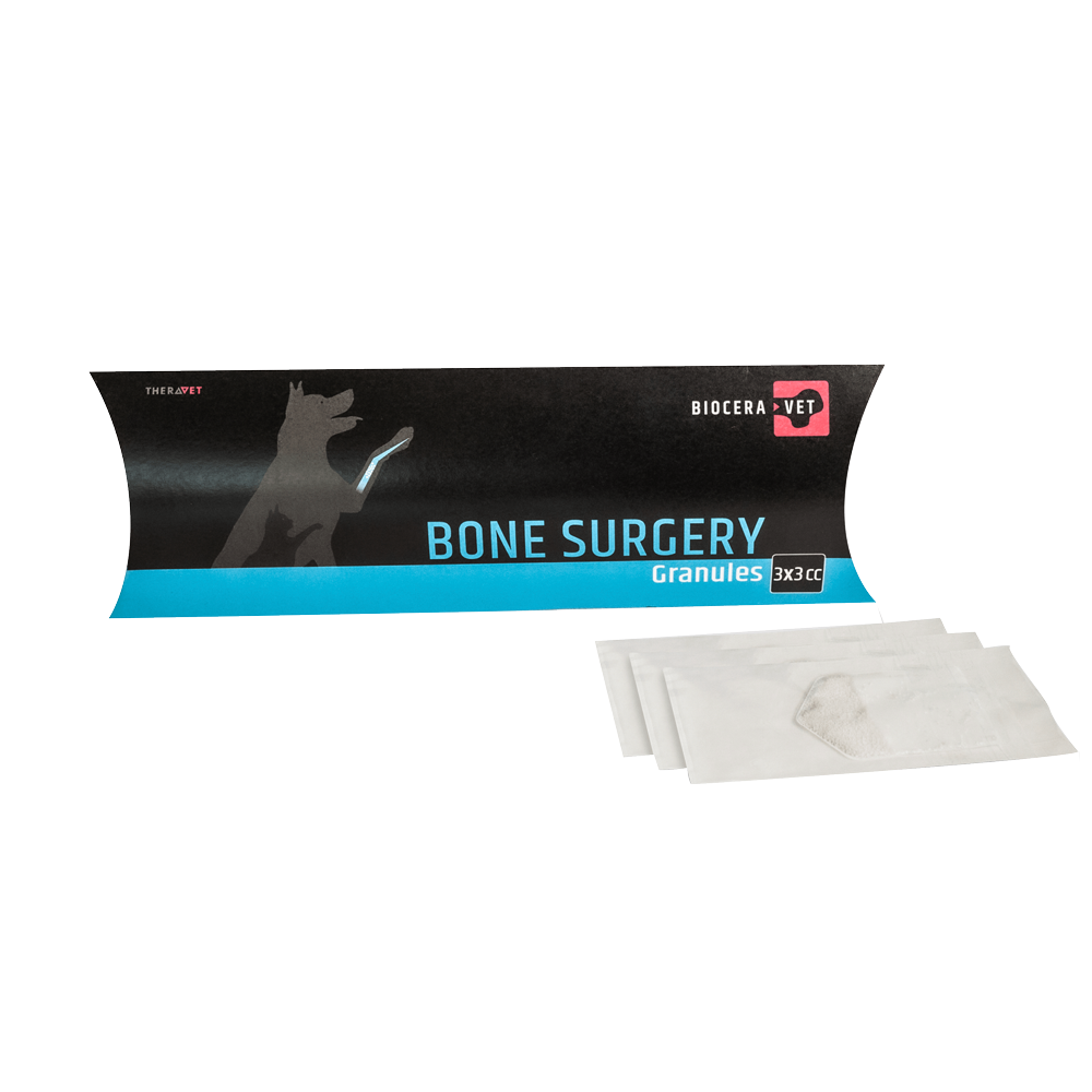
A Dachshund dog, “Wombat”, presented in February for treatment of a furcation (F2) exposure and increased periodontal probing depths (7 – 9mm on the buccal surface) of 309. Dental radiographs using a Sopix2 digital sensor showed horizontal alveolar bone loss. Treatment of the furcation involved elevating a gingival envelope flap and open root planing, followed by placement of an alloplast comprising of hydoxyapatite and beta tricalcium phosphate into the defect. Sanos was applied post treatment.
Step by step

Presentation
A: Photograph of the left side of Wombat’s mouth demonstrating increased plaque and calculus accumulation. Note: due to loss of the caudal maxillary teeth, 309 has plaque build-up and gingival inflammation.

Envelope flap & root plane
B: Photograph 309 after envelope flap and root planning was completed. Note: furcation exposure and radiograph sensor placed lingually.

Initial radiograph
C: Radiograph 309 showing horizontal bone loss and furcation exposure.

Placement of bone graft granules
D: Photograph showing placement of bone graft granules into 309 furcation defect.

Presentation
A: Photograph of the left side of Wombat’s mouth demonstrating increased plaque and calculus accumulation. Note: due to loss of the caudal maxillary teeth, 309 has plaque build-up and gingival inflammation.

Envelope flap & root plane
B: Photograph 309 after envelope flap and root planning was completed. Note: furcation exposure and radiograph sensor placed lingually.
Six month follow up

Presentation
G: Photograph at 6 month follow-up showing left side of Wombat’s mouth demonstrating normal gingival height with minimal inflammation and no calculus accumulation.

6 month post-treatment radiographs
H & I : Radiographs of 309, 6 months post placement of bone graft, demonstrated new bone formation within the furcation defect and some increase in alveolar bone height adjacent to the furcation.


